


 824 844

[(Fig._2)TD$FIG]
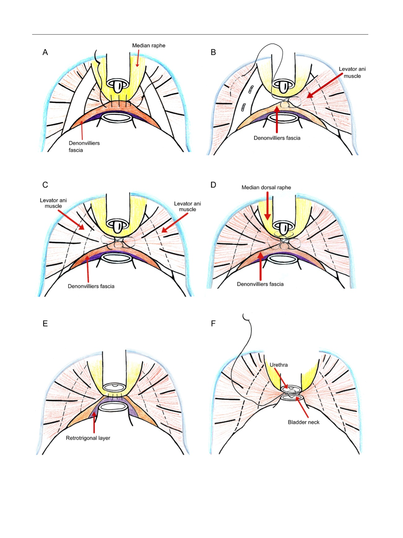
Fig. 2 – (A) Control group. Suture of the Denonvilliers fascia and bladder to the median dorsal raphe according to Rocco. (B) In the intervention group,
we used an absorbable monofilament barbed V-loc 2/0 suture, which we led first to the right, across the medial levator ani muscle, then through the
Denonvilliers fascia without injuring the neurovascular bundles. (C) The stitch goes over the bundles of the left medial levator ani and back to the
Denonvilliers fascia. (D) The end of the stitch is passed under the urethra through the median dorsal raphe. (E) The suture is passed back through the
detrusor under the bladder neck through the retrotrigonal layer. (F) In the last step, the needle is passed through the bladder neck and urethra to
align them.
E U R O P E A N U R O L O G Y 7 1 ( 2 0 1 7 ) 8 2 2 – 8 3 0
824