


 795 844

Symptom Score [IPPS]) to assess the severity of benign
prostatic hyperplasia. It was subsequently demonstrated
that this questionnaire is accurate in describing LUTS in
women
[51]. Fedele et al
[44]modified the AUASI/IPPS,
replacing three questions concerning obstructive symp-
toms with three questions concerning irritative symptoms,
especially during the perimenstrual period; this modified
questionnaire allows evaluation of the presence of specific
catamenial symptoms related to BE in patients with high
suspicion for this disease. The questionnaire was adminis-
tered to 154 patients undergoing surgery for CPP. Of the
total study population, 127 (82.4%) patients had pelvic
endometriosis and 14 (9.0%) had BE. The questionnaire was
effective in identifying BE (area under the receiver
operating characteristic curve 0.951)
[44].
More recently, Ballester et al conducted two studies
investigating the presence of urinary dysfunction in
patients with DIE before
[36,52]and after surgery
[36]using the IPPS
[52]and the Bristol Female LUTS (BFLUTS)
questionnaire
[36,52]. The BFLUTS questionnaire comprises
three domains: symptom questions (frequency of micturi-
tion, nocturia, urgency, urge incontinence, bladder pain,
frequency incontinence, stress incontinence, unpredictable
miscellaneous incontinence, volume of leakage, hesitancy,
strain to start, intermittency, nocturnal incontinence,
reduced stream, acute retention, burning, incomplete
emptying, stopping flow, and frequency between voiding)
mostly with a corresponding subquestion; sexual function
questions (pain due to dry vagina, disturbed sex life, pain
during intercourse, and leakage during intercourse); and
quality-of-life questions (change of underwear/use of pads,
number of changes, change of outer clothing, reduction in
fluid intake, affected daily tasks, avoidance of situations
where no toilet is available, interference with physical
activity, interference with social life, overall interference
with life, how long symptoms have been bothering, and
notion of spending rest of life with no change).
In conclusion, the use of validated questionnaires may be
helpful in the management of BE. In particular, the modified
AUASI questionnaire by Fedele et al should be utilized
during the diagnostic work-up to improve the detection of
BE. The IPPS and the BFLUTS questionnaires are useful in
assessing the variegated spectrum of LUTS associated with
DIE, including BE, and in monitoring changes in symptoms
after treatment. However, administration and answering of
these questionnaires are associated with significant time
costs in routine clinical practice, so we recommend their use
mainly for scientific research purposes
( Table 1).
3.2.4.
Ultrasonography
Ultrasonography is fundamental in the diagnosis of BE and
in planning the most appropriate treatment, since it can be
used to evaluate the location and size of the nodule, and to
estimate the distance between the lesion borders and the
ureteral orifices
( Table 1)
[53] .The first description of the
sonographic features of BE was provided in 1980 by
Goodman and colleagues
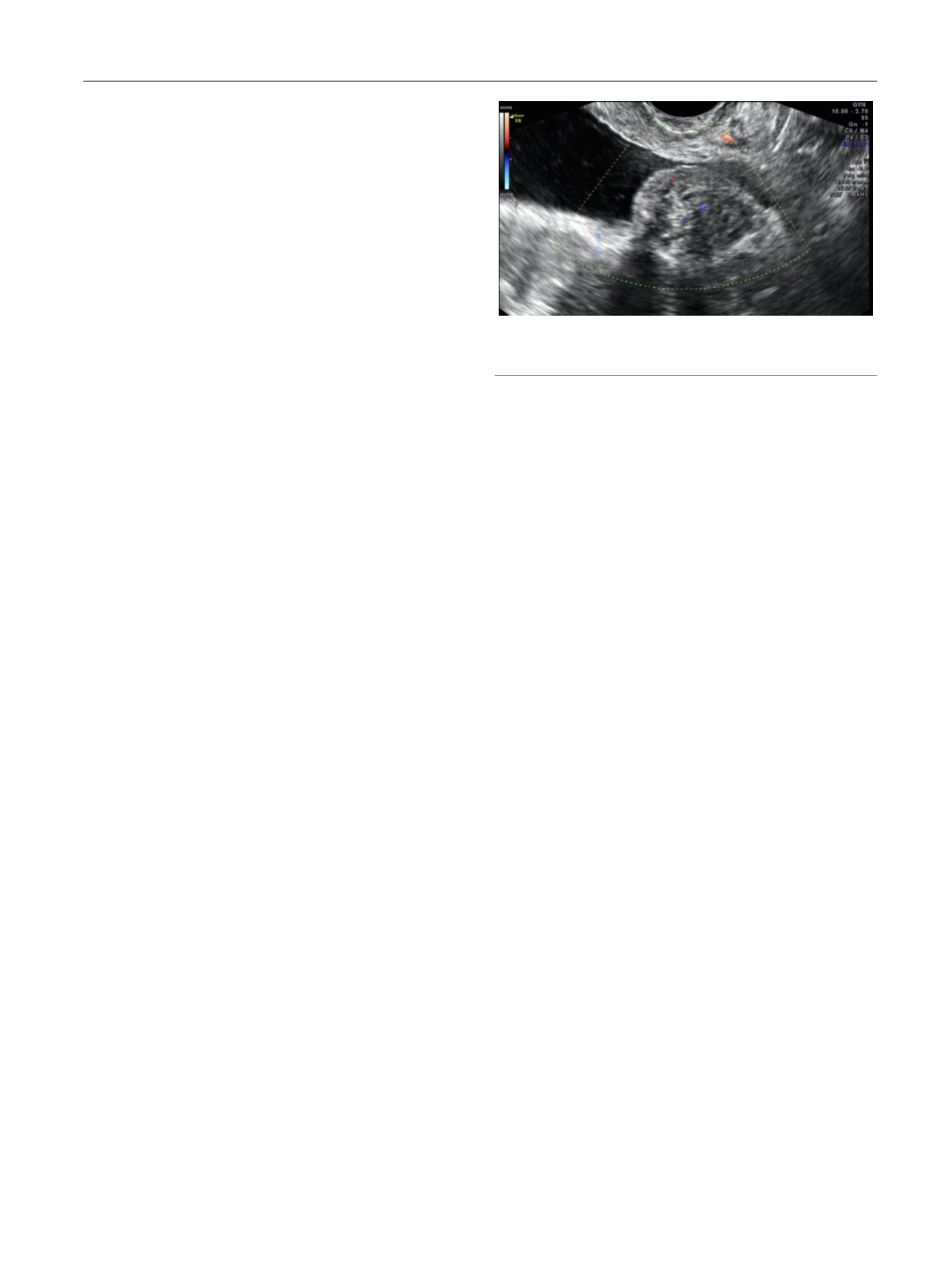
[54]. On ultrasonography with
the bladder full of anechoic urine, BE appears as a filling
defect of the posterior wall with a variable protrusion into
the lumen, with an iso/hypoechoic aspect sometimes visible
with small transonic formations that are usually not
vascularized
( Fig. 2). Bladder nodules are usually spherical
or comma-shaped with regular contours, but sometimes the
lesion borders can be irregular, raising a suspicion of
malignancy. However, bladder nodules are usually covered
by a small rim of the hyperechogenic layers of the bladder
wall (submucosal and serosa), while spiky or papillary
projections interrupt the hyperechogenic layers of the
bladder wall in cancer. Color Doppler may physicians in
establishing a differential diagnosis, since it commonly
reveals minimal to moderate internal blood flow in patients
with BE
[55,56].
In 1997, an Italian study including six patients compared
abdominal ultrasonography, transvaginal ultrasonography
(TVS), and MRI in the preoperative evaluation of BE. All the
techniques identified bladder lesions; however, TVS was the
most accurate in defining the size of the lesions, infiltration
of the detrusor muscle, and continuity with extravesical
lesions
[57] .Both abdominal ultrasonography and TVS may
be used to detect vesical endometriotic lesions; however, in
gynecologic clinical practice, TVS is the preferred technique.
Table 1summarizes the results of studies investigating the
use of TVS to diagnose BE
[40,41,58–64]. A recent
systematic review and meta-analysis revealed overall
pooled sensitivity of 62% (95% CI, 40–80%), specificity of
100% (95% CI, 97–100%), positive likelihood ratio of 208.4
(95% CI, 21.0–2066.0), and negative likelihood ratio of 0.38
(95%CI, 0.22–0.66) for TVS detection of BE. The study
suggests that TVS is a useful first-line method for
diagnosing BE in clinical practice. The pretest probability
was 5%, which increased to 92% when suspicion of DIE was
present after TVS examination, and fell to 2% in the absence
of ultrasonographic findings in the bladder
[65]. Tammaa
et al
[64]evaluated interobserver agreement and accuracy
for TVS in diagnosing endometriomas and DIE, and also
considered BE. Patients were independently examined
prospectively by two experienced sonographers who were
blinded to the other’s results; Gwet’s first-order agreement
coefficient (Gwet’s AC1) was used to calculate interobserver
agreement. The study demonstrated that TVS had high
accuracy and specificity but fair sensitivity in the diagnosis
of BE
( Table 2 ); most importantly, TVS was highly
[(Fig._2)TD$FIG]
Fig. 2 – Endometriotic bladder nodule on transvaginal ultrasonography.
Color Doppler reveals no vascularization.
E U R O P E A N U R O L O G Y 7 1 ( 2 0 1 7 ) 7 9 0 – 8 0 7
795