


 782 844

bone metastases but preserved in a subgroup of cases.
Notably,
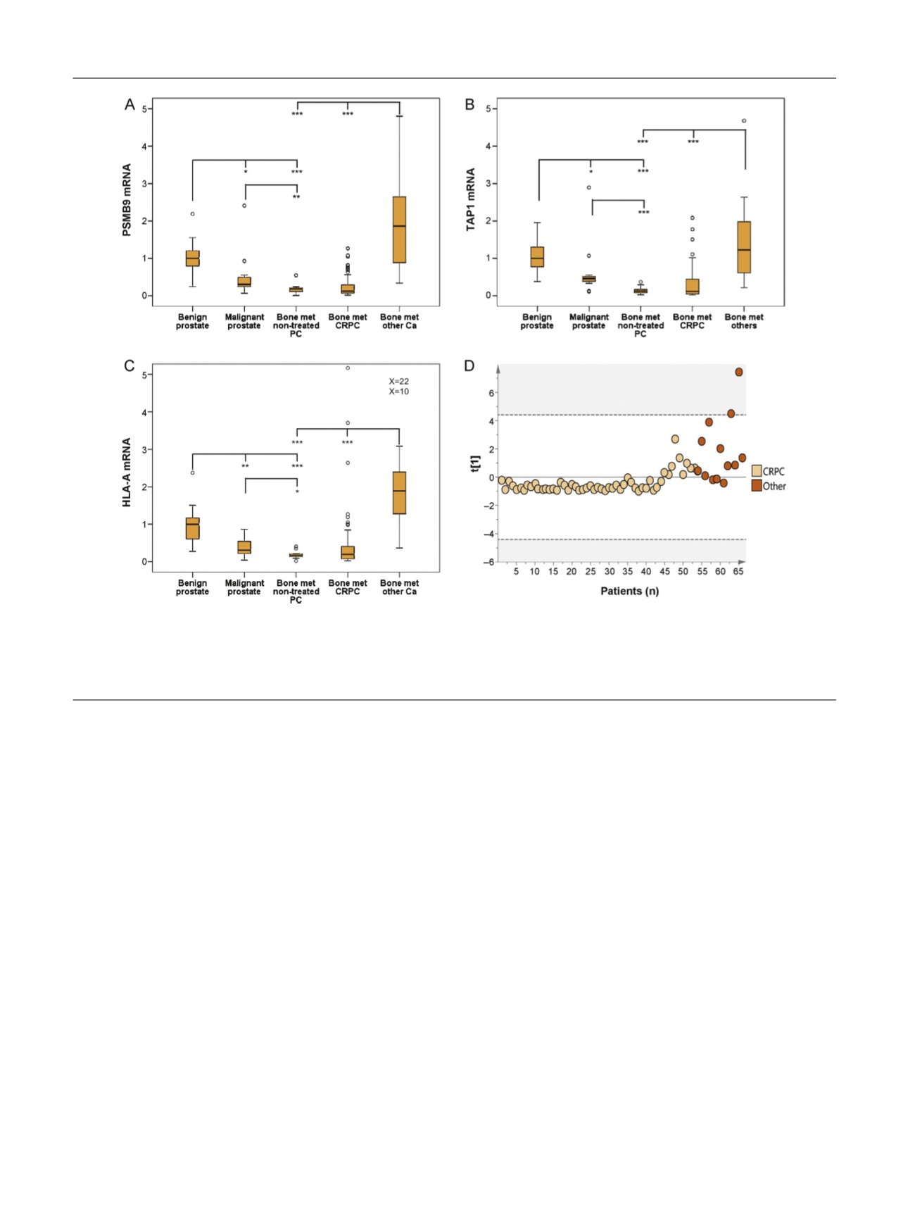
PSMB9
,
TAP1
, and
HLA-A
mRNA levels were all
significantly lower in malignant compared to nonmalignant
prostate tissue and were even lower in bone metastasis
tissue
( Fig. 3 A–C).
Accordingly, immunoreactivity for HLA class I ABC was
lower in metastases than in matched primary tumor
biopsies obtained at diagnosis (median 37 mo [IQR 16–79
mo] before metastasis surgery;
p
= 0.037,
n
= 29; data not
shown), indicating a reduction in MHC class I protein
expression during PC disease progression.
3.4.
Inverse correlation between MHC class I expression and
nuclear AR immunoreactivity in CRPC metastases
The PCA model indicated downregulated MHC class I
antigen presentation in AR-driven CRPC bone metastases
compared to preservation of MHC class I antigen presenta-
tion in non–AR-driven CRPC bone metastases, so we studied
HLA class I ABC immunoreactivity in relation to nuclear AR
immunoreactivity (previously measured in those metasta-
ses and assumed to reflect AR activity
[6] ). HLA class I ABC
immunoreactivity in tumor cells was evaluated in metasta-
ses for which FFPE tissue was available, and was found to be
inversely correlated to the nuclear AR score (Rs = 0.49,
p
= 0.001,
n
= 41;
Fig. 4A–D). Importantly, metastases with
moderate to intense HLA class I ABC immunoreactivity
showed a significantly higher frequency of CD3
+
infiltrating
cells than cases with negative to weak immunostaining
(Supplementary Fig. 3).
Staining heterogeneity was observed for both AR and
HLA class I ABC immunoreactivity in many cases, so double
staining was performed. Nuclear AR showed a clear inverse
staining pattern to HLA class I ABC
( Fig. 4E). FOXA1 staining
in consecutive sections indicated reduced but not complete-
ly diminished FOXA1 levels in AR-negative/HLA class I ABC–
positive tumor cells
( Fig. 4 F).
3.5.
Reduced expression of HLA class I ABC in primary prostate
tumors with advanced disease stage
To evaluate if MHC class I expression in primary PC is
related to patient prognosis, HLA class I ABC immunoreac-
tivity was evaluated in a TMA including transurethral
cancer biopsies from 284 patients with long clinical follow-
up and in adjacent benign tissue in 179 cases. Malignant
epithelial cells showed less intense staining than adjacent
benign epithelial cells (
p
<
0.0001;
Fig. 5 A). Lower HLA ABC
[(Fig._3)TD$FIG]
Fig. 3 – Relative mRNA levels of (A)
PSMB9
, (B)
TAP1
, and (C)
HLA-A
in paired nonmalignant and malignant prostate tissue samples from patients
treated with radical prostatectomy (
n
= 12) and in non-treated (
n
= 11) and castration-resistant prostate cancer (CRPC) bone metastases (
n
= 53) and
bone metastases from other malignancies (
n
= 13). *
p
< 0.05, **
p
< 0.01, ***
p
< 0.001. (D) Principal component analysis of bone metastases samples
from CRPC patients (beige) and patients with other malignancies (
[6_TD$DIFF]
orange). Score plot for the first principal component, for which each dot
corresponds to one patient sample. Samples cluster according to their relative
PSMB9
,
TAP1
, and
HLA-A
mRNA levels. met = metastases.
E U R O P E A N U R O L O G Y 7 1 ( 2 0 1 7 ) 7 7 6 – 7 8 7
782